A 45-year-old man presents to the emergency department for evaluation of a “fluttering heart,” dizziness, and shortness of breath, which have been constant since yesterday evening. Past medical history is significant for atrial fibrillation and hypertension. Of note, the patient typically takes daily metoprolol, but he was unable to afford his most recent prescription refill. Physical examination is significant for a blood pressure of 90/50 mm Hg, heart rate 122 beats/minute, and an irregularly irregular heartbeat. A normal jugular venous pressure (JVP) tracing is pictured below.
Which of the following changes to the jugular venous pressure (JVP) tracing is most expected in this patient?
A) Attenuated A wave and X descent
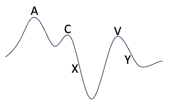
This patient is likely in atrial fibrillation, which attenuates atrial systole and atrial kick (represented by the A wave). Because the right atrium fails to fully empty into the right ventricle, the X descent that typically results from ventricular systole, relieving pressure on the right atrium, cannot occur.
Answer choice B: Attenuated X descent with prominent V wave, is incorrect. This JVP wave form occurs with tricuspid regurgitation. The V wave, which occurs towards the end of ventricular systole and maximal atrial filling, is prominent due to back flow of blood into the right atrium through the tricuspid valve.
Answer choice C: Attenuated Y descent only, is incorrect. This JVP wave form occurs with cardiac tamponade, which impairs ventricular diastolic filling.
Answer choice D: Prominent A wave only, is incorrect. This JVP wave form occurs with atrio-ventricular (AV) dissociation and is also known as a “cannon A wave.” It occurs due to contraction of the right atrium against a closed tricuspid valve.
Answer choice E: Prominent A wave with attenuated Y descent, is incorrect. This JVP wave form occurs with tricuspid stenosis. The stenotic valve attenuates flow of blood from the right atrium to right ventricle during ventricular diastole.
Key Learning Point
Jugular venous pressure tracings reflect cardiovascular pathology and are composed of 5 main components: an A wave representing atrial contraction, a C wave representing the beginning of ventricular systole/the bulging and closure of the tricuspid valve back into the right atrium. The x descent in the JVP is due to atrial relaxation following the summit of the "a" wave formed by atrial contraction during sinus rhythm. The x descent occurs during ventricular systole with rising RV pressure. The V wave represents maximal atrial filling, and a Y descent representing the beginning of ventricular diastole.